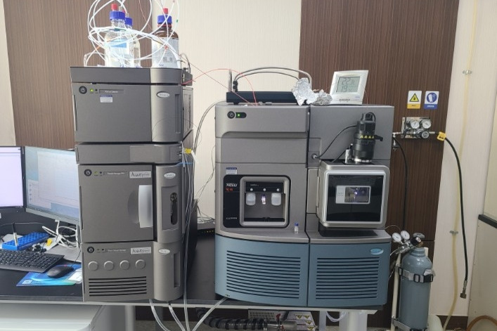
Device built-in
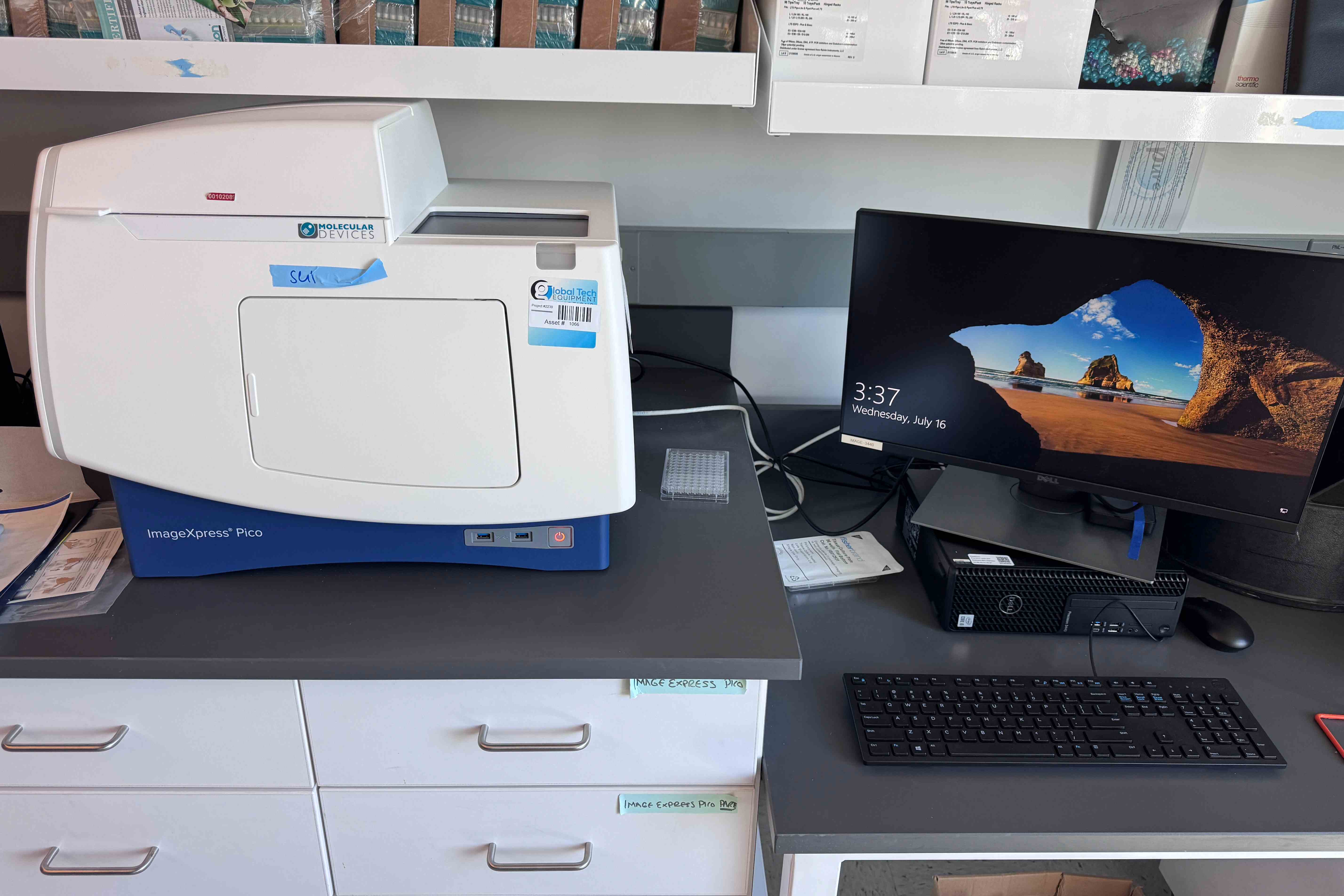
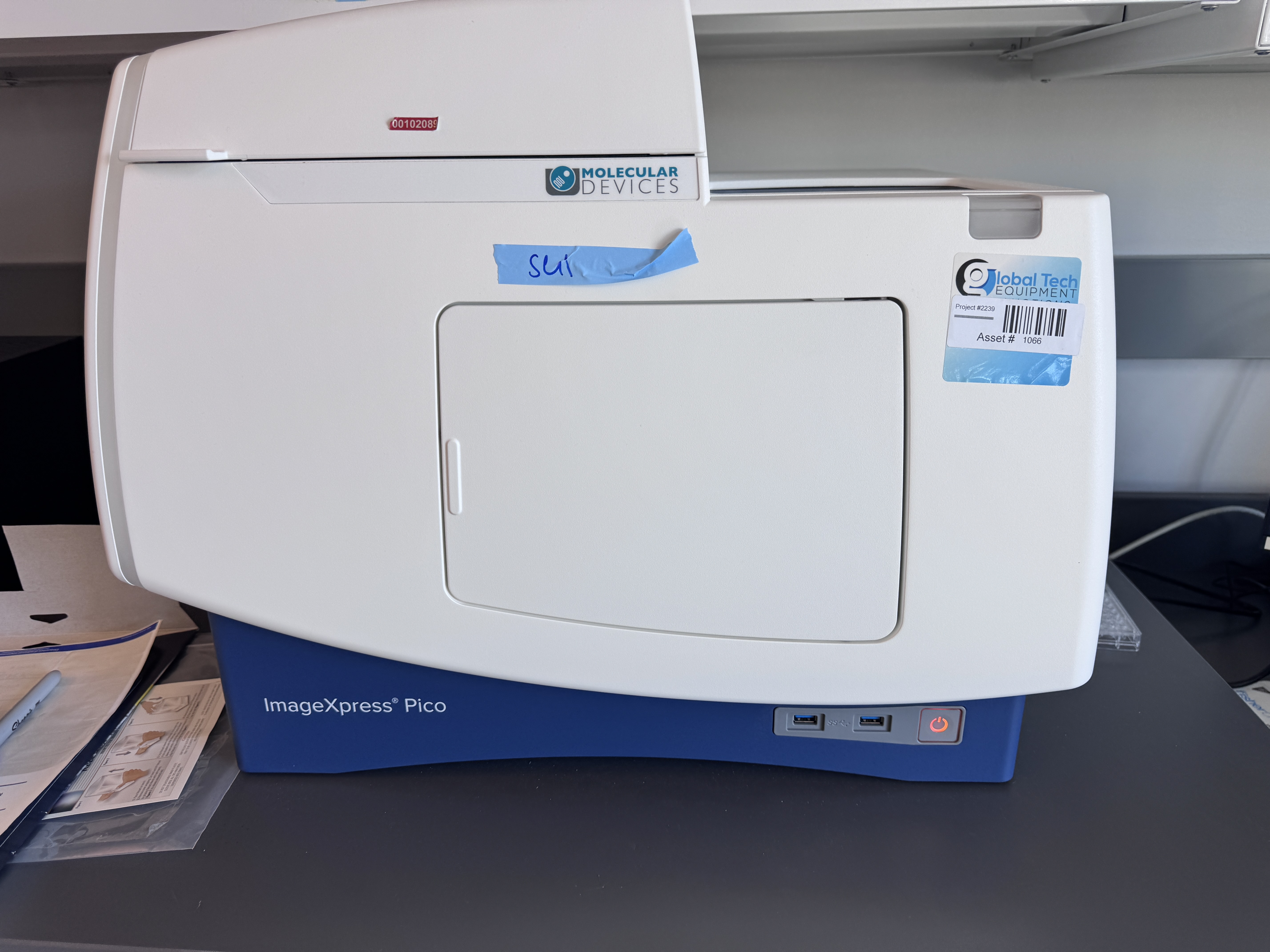
Molecular Devices ImageXpress Pico Automated Cell Imaging System: Bringing High-Content Analysis Power to Every Lab
In modern life sciences research, particularly in drug discovery and cell biology, the precise and efficient quantitative analysis of cellular phenotypes has become crucial. High-content screening (HCS) technology, which perfectly integrates automated microscopy with multi-parameter image analysis, is a core tool to meet this challenge. However, traditional HCS systems are often large, complex, and costly, putting them out of reach for many laboratories. Molecular Devices (often referred to as "美谷分子" in China) addressed this need by introducing the ImageXpress Pico Personal High-Content Imaging System, designed to bring high-performance automated imaging and analysis capability to the benchtop, making it a powerful tool easily accessible to every laboratory.
I. Core Positioning: Desktop High Performance, Ushering in the Era of Personal HCS
The core value of the ImageXpress Pico lies in its "personal" nature and "ease of use." It is not a simplified version of a traditional large HCS system but a thoughtfully designed, integrated solution. Its compact footprint allows it to fit easily inside a standard laminar flow hood or biosafety cabinet for dynamic live-cell monitoring under sterile conditions. The system integrates the computer, control software, and user interface, offering "turnkey" operation. This design enables even small-to-midsize labs, satellite labs of core facilities, or research groups focused on specific projects without a dedicated operator to independently conduct high-quality cell imaging and quantitative analysis, significantly enhancing research flexibility and efficiency.
II. Technical Features and Performance Advantages
Excellent Optical Imaging Quality:
High-Precision Automation: The system features fully automated components including an objective turret, filter cube wheel, stage, and focus mechanism, ensuring fast, accurate, and reproducible imaging.
Superior Image Quality: Utilizes a scientific-grade sCMOS camera combined with high-quality optics and LED illumination to capture high-sensitivity, low-noise, sharp images, clearly presenting both bright fluorescent signals and weak autofluorescence.
Intelligent Focusing: Incorporates multiple focusing modes like laser autofocus and software autofocus, enabling rapid and precise identification of the focal plane, which is essential for multi-well plate time-course experiments, ensuring data consistency.
Integrated Intelligent Analysis and Software:
Powerful Analysis Software: Comes pre-installed with robust MetaFlux™ or CellReporter™ analysis software. The software includes optimized, standardized application modules for "one-click" analysis of complex phenotypes like cell counting, protein expression quantification, nuclear translocation, neurite outgrowth, 3D spheroids, and stem cell colony formation.
Wizard-Based Interface: The software interface is intuitive and user-friendly, providing step-by-step guides for experiment setup, image acquisition, and quantitative analysis, significantly reducing the learning curve.
Comprehensive Data Management: The software not only generates detailed quantitative data but also automatically creates visualizations like scatter plots and heat maps, and supports comprehensive report generation, helping researchers deeply explore the biological meaning behind the data.
Flexible Application Compatibility:
The system supports various labware, including 6- to 384-well plates, dishes, and slides, meeting the needs of different throughput experiments.
It is suitable for endpoint assays and is particularly ideal for multi-day live-cell imaging, making it a perfect platform for studying cell proliferation, apoptosis, migration, and toxicity.
III. Broad Application Scenarios
The application range of the ImageXpress Pico is extremely wide, covering almost all areas requiring quantitative cell analysis:
Drug Discovery: Used for high-content screening of small molecule compounds or siRNA libraries to assess their effects on cell morphology, proliferation, toxicity, and more.
Cell Biology Research: Quantitatively studies key life processes like cell cycle, apoptosis, autophagy, receptor internalization, and signal transduction.
Neuroscience: Automatically quantifies neuronal morphology, neurite length, and branching complexity.
Virology & Infection: Monitors viral infection processes and their effects on host cells.
Stem Cell Research: Used for stem cell colony formation analysis, pluripotency identification, and differentiation monitoring.
Toxicology Studies: Assesses the potential toxic effects of compounds on cells.
IV. Summary
The Molecular Devices ImageXpress Pico system successfully breaks down the technical barriers to high-content analysis. It skillfully strikes an optimal balance between performance, ease of use, and footprint, transforming the powerful analytical capability once available only in large core facilities into a routine tool that any researcher can easily operate on their own bench. It is not just a high-performance imager but a complete, integrated solution for image acquisition and intelligent analysis. For life science laboratories committed to enhancing research quality and accelerating scientific progress, the ImageXpress Pico is undoubtedly a "personal research assistant" that significantly boosts core competitiveness.