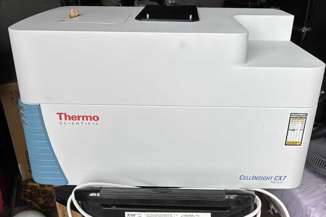
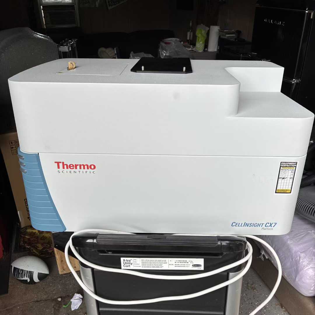
Thermo Fisher CellInsight CX7 High-Content Analysis Platform
The Thermo Fisher CellInsight CX7 High-Content Analysis Platform is an advanced research instrument integrating automated microscopic imaging and intelligent data analysis, specifically designed for life science research and drug screening. It offers high-throughput, high-precision, and multi-modal imaging capabilities. Below is a detailed introduction covering its core technology, functional applications, and key advantages:
Price:Negotiable
Service charge: 10% of the transaction price
- Equipment brand:Thermo Fisher
- Equipment installation time:2023
- Equipment location:Shanghai
- Equipment model:Thermo Fisher CellInsight CX7 High-Content Analysis Platform
- Equipment serial number:
- Voltage:
- Electrical frequency:
- Size:
- Weight:
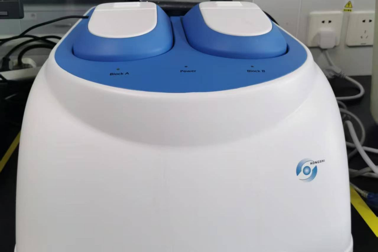
Device built-in
Thermo Fisher CellInsight CX7 High-Content Analysis Platform
The Thermo Fisher CellInsight CX7 High-Content Analysis Platform is an advanced research instrument integrating automated microscopic imaging and intelligent data analysis, specifically designed for life science research and drug screening. It offers high-throughput, high-precision, and multi-modal imaging capabilities. Below is a detailed introduction covering its core technology, functional applications, and key advantages:
I. Core Technology
Multi-Modal Imaging System
The CellInsight CX7 supports brightfield, widefield fluorescence, and confocal imaging modes, equipped with 7 fluorescence channels (covering UV to near-infrared spectra) and 5 brightfield channels. Its laser-based autofocus technology enables rapid sample positioning, ensuring imaging stability even with irregularly distributed wells. The confocal module employs dual-pinhole Nipkow disk technology (switchable between 40μm/70μm), significantly enhancing resolution for thick samples (e.g., 3D organoids). The light source uses solid-state LED or laser engines (LZR Pro version), with a lifespan exceeding 20,000 hours and an excitation wavelength range of 386nm to 740nm, meeting the needs of multiplex fluorescence labeling.
High-Performance Hardware Configuration
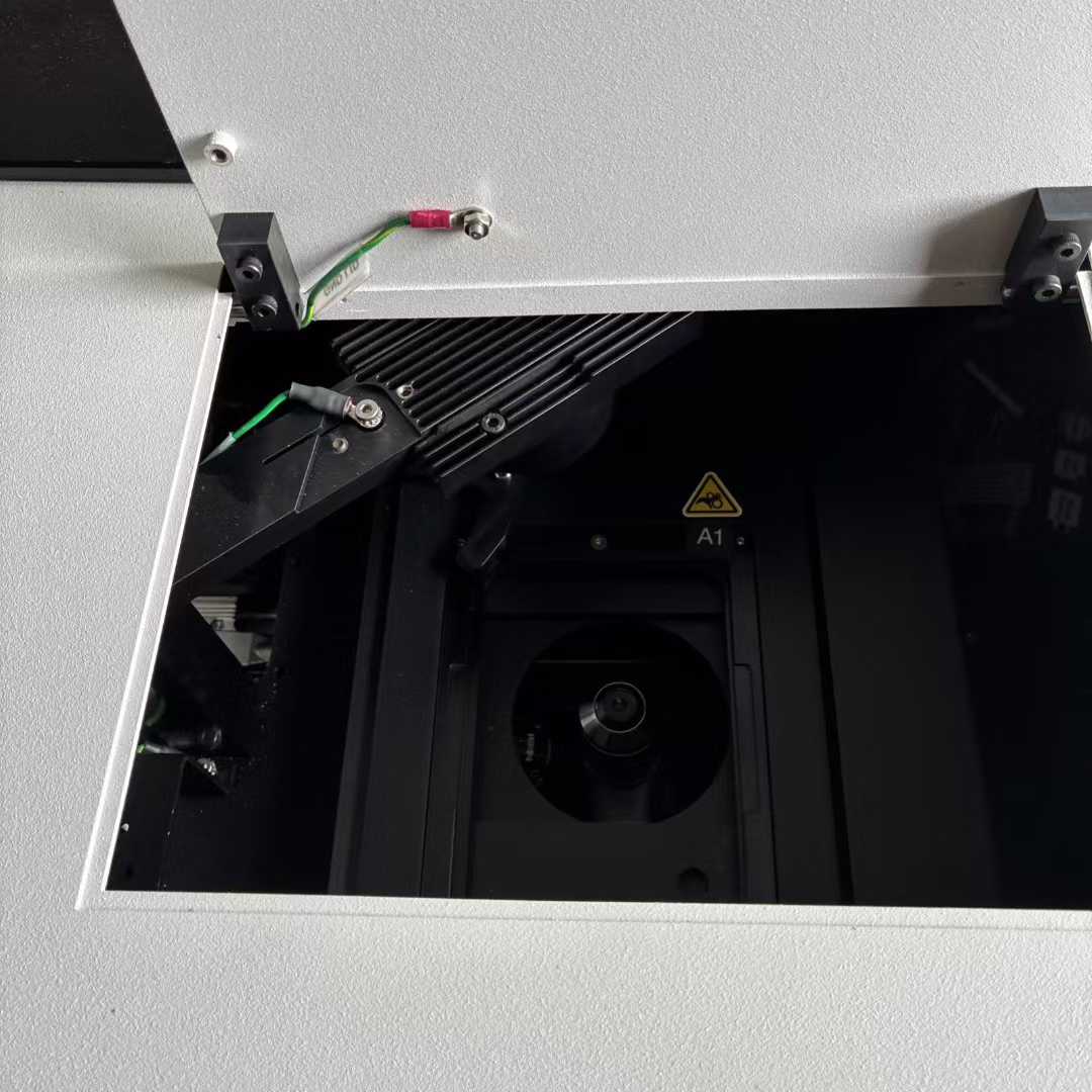
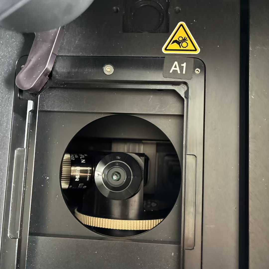
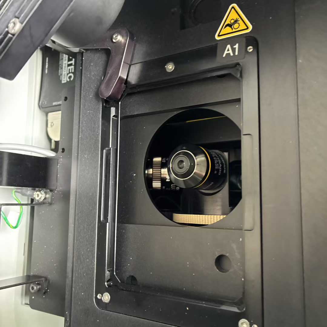
The platform features a back-illuminated sCMOS camera with ≥95% quantum efficiency, supporting -20°C cooling and 65,536 grayscale levels, significantly reducing background noise and improving dynamic range. Olympus X-line objectives (standard 10X-40X, optional 60X) further optimize image quality, particularly for publication-grade imaging. Z-axis precision reaches 25nm, and XY-axis precision is 50nm, ensuring accurate capture of ultra-fine structures.
Intelligent Analysis Software
The built-in HCS Studio 5.0 software provides over 40 preset analysis modules (e.g., apoptosis, autophagy, cell cycle) and supports custom algorithm development. The software delivers real-time data feedback; for example, analyzing neural axon growth in a 96-well plate takes only 4 minutes, with automatic generation of statistical results. Its unique EurekaScan Finder function dynamically tracks spheroids or rare cellular events.
II. Functional Applications
Basic and Translational Research
The platform is suitable for studying various biological processes:
Angiogenesis: Automatically quantifies microvessel count and branching index;
Apoptosis Detection: Analyzes DNA damage via TUNEL labeling or caspase activity;
Autophagy Analysis: Quantifies LC3B protein vesicle-based immunofluorescence;
Cell Cycle: Evaluates mitotic index via DNA content or phosphorylated histone H3 staining.
Drug Screening and Toxicology
Supports high-throughput screening for 6- to 1536-well plates, compatible with SBS-standard microplates and tissue slides. For example, it quantifies neutral lipid droplet accumulation in lipotoxicity assays or analyzes dose effects of cell proliferation inhibitors via EdU incorporation.
Live-Cell and 3D Model Studies
Optional environmental control modules (temperature, humidity, CO₂/O₂ regulation) enable long-term live-cell observation. Its confocal mode is particularly suited for tomographic imaging of 3D cell spheroids and organoids, providing critical tools for oncology and developmental biology.
III. Key Advantages
Efficiency and Flexibility
The intelligent acQuisition (iQ) technology enables "scan while analyzing," shortening experimental cycles. Users can freely switch imaging modes, such as combining widefield (rapid screening) and confocal (high-resolution details) in the same experiment.
Scalability and Compatibility
The platform can connect to automated robotic arms (e.g., Orbitor RS processor) to increase throughput and seamlessly integrates with Molecular Probes fluorescent reagents. The Store Express database software supports multi-user remote data management.
User-Friendly Design
Suitable for both beginners and advanced users: preset analysis templates simplify operations, while advanced tools support complex parameter adjustments like background correction and ROI segmentation.
Summary
The CellInsight CX7 series (including Pro and LZR Pro versions), with its modular design, multi-modal imaging, and powerful analysis capabilities, has become a core instrument in fields such as cell biology and drug development. Its price ranges approximately from 1.5 to 3.5 million RMB (depending on configuration), and some models are available through tender procurement. For further technical specifications or application cases, refer to Thermo Fisher’s official resources or contact local distributors.